Electrophysiology Study
What is a Cardiac Electrophysiology Study?
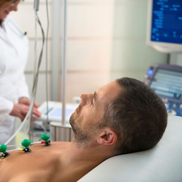
A cardiac electrophysiology study is a study performed by a Heart Specialist with specialized training in abnormal rhythms of the heart. To have this study performed you would need to be admitted to a hospital with specialized equipment necessary to perform this procedure. You will generally be admitted on the day of the procedure and will need to fast for 3-4 hours prior to the procedure.
How is it done?
The procedure is performed using local anaesthesia with IV sedation which will be given in doses requisite to make you feel calm and slightly sleepy but you will not be rendered unconscious. Small plastic tubes will be placed in the veins of the leg or arm. Through these tubes insulated wires or catheters are placed through the veins into the heart. These catheters are then connected to very sensitive high fidelity recording equipment which will record the electrical signal as it passes through the heart.
The normal conduction pathway will then assessed and a search made for any abnormal conduction pathways that may be present. Using very minute electrical charges we can stimulate the heart to beat rapidly or slowly and we will try to induce any rapid palpitations that will help identify the mechanism of the symptoms that you have been experiencing. Generally we can stop and start the heart at will using these electrical stimuli.
The procedure yields very useful information in patients with abnormal heart rhythms and for patients with recognized circuits causing rapid palpitations are readily identified and we can then proceed to a curative procedure with high success , resulting in cure rates in the order of 90-95%.
What are the risks?
The risks of the procedure include puncturing the heart with the catheters 1:1000 which can lead to bleeding around the heart which may need to have the chest opened to put a stitch in the bleeding point. Additionally there is always a risk of bruising, bleeding and thrombosis in the leg in which the catheters have been inserted. You are usually given a blood thinner to reduce the risk of this happening. Should you develop a thrombosis in your leg it may travel through the circulation to your lungs and cause difficulty with breathing. The catheters are usually advanced using Xray guidance thus there will be moderate radiation exposure. This is particularly relevant to patients who may be pregnant and generally we can use the newer 3D mapping systems which use electroanatomic sensors to detect the catheters.
However, 99% of patients have the procedure performed without any major risk and can be discharged home the following day.
If you have any further questions, please contact us at:
Queensland Cardiology
St Vincent’s Private Hospital Northside
North Medical Suites, Green Lifts Level 3,
627 Rode Road
Chermside Q 4032
(07) 3861 5522

