Endomyocardial Biopsy
What is an Endomyocardial Biopsy?
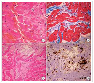
An endomyocardial biopsy is a procedure in which your doctor takes samples of heart tissue to help diagnose a number of cardiac conditions.
How is it done?
The procedure begins with an injection of local anaesthetic into your right groin. A fine tube (catheter) is put into the vein in the groin. You may feel pressure in your leg while the tube is placed in the vein. The tube is passed up until it reaches the right heart chamber. This is painless. The doctor will use X-Ray images to see and guide the catheter.
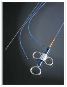
‘Biopsy forceps’ or pincers at the end of the catheter, are used to take the tissue samples. This is repeated at least 4 times to get enough samples. The biopsy pieces are very small (one to two millimetres in diameter). You will feel a few extra heart beats, otherwise this part of the procedure is also painless.
At the end of the procedure the forceps and catheter are removed.
How do I prepare?

No food or drink 3 hours prior to the procedure


Biopsy Forceps

After the procedure
- You may have some bruising and soreness around the access site
- Do not drive for 24 hours after the procedure
- Do not lift objects over 5kg for 3-4 days after your procedure
- Notify your Doctor or present at the emergency department if you have any significant bleeding, pain or fevers
- Notify your Doctor if you have any significant chest pain or angina
What are the risks?
In recommending this procedure, your doctor has balanced the benefits and risks of the procedure against the benefits and risks of not proceeding. Your doctor believes there is a net benefit to you going ahead. This is a very complicated assessment.
Common risks (more than 5%) –
- minor bleeding and bruising at the puncture site
- abnormal heartbeat lasting several seconds which settles by itself
Uncommon risks (1-5%) –
- Unable to get the catheter into the leg vein. The procedure may be changed to the opposite leg or to a different approach e.g. the neck or an arm vein
- abnormal heart rhythm that continues for a long time. This may need an electric shock to correct.
- the femoral artery (in the groin) is accidentally punctured. This usually just requires pressure on the artery. However, rarely this may require surgery to repair.
- unable to get any heart samples. This may be due to scarring of the heart.
Rare risks (less than 1%) –
- infection. This will need antibiotics.
- allergic reaction to the local anaesthetic. This may require some medication to treat.
- embolism. A blood clot may form and break off from the catheter. This is treated with blood thinning medication.
- a hole is accidentally made in the heart or the heart valve. This will need surgery to repair.
- damage to the nerve in the leg.
- air embolism. Oxygen may be given.
- a stroke. This may cause long term disability.
- death as a result of this procedure is extremely rare.
If you have any further questions, please contact us at:
Queensland Cardiology
St Vincent’s Private Hospital Northside
North Medical Suites, Green Lifts Level 3,
627 Rode Road
Chermside Q 4032
(07) 3861 5522

