The Normal Heart
The heart structure has 6 components:
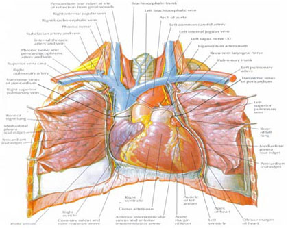
1. Heart muscle, which pumps the blood around the body
2. Electrical system, which generates and stimulates the heart muscle to pump
3. A circulatory system to supply itself with blood
4. Heart valves, four sets which ensures that the blood keeps moving forward
5. A fibrous skeleton to which the muscle and valves are attached
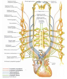
6. Nerve supply which allows the heart to respond to stress and emotion
Heart Muscle
The average heart beats 70 times per minute which is on average 100,000 times per day or 36,500,000 times per year. Most people are generally unaware of the heart beating except during periods of high emotion or physical exertion. The heart pumps on average 5L of blood around the system every minute and this can be increased 5-6-fold during periods of peak exertion. The normal heart ejects on average 55% of its volume with each beat and with exertion becomes much more vigorous and can inject up to 80% of its volume with each beat. Your whole blood volume circulates through your vascular system every 10 seconds.
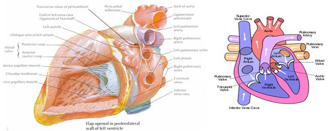
The Electrical System
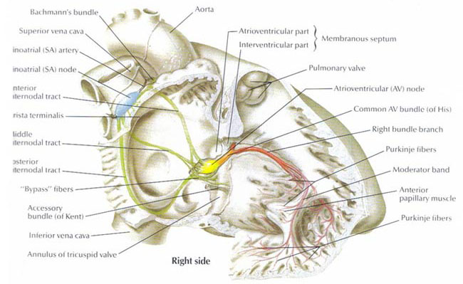
The heart beats independently of the nervous system and will continue to beat even if removed from the body so long as nutrients and oxygen are supplied. The signal is transferred through the atrial muscles very rapidly and this causes them to beat uniformly. The signal then collects again at a relay station, the Atrio-Ventricular (AV) node, which is another area of specialized conduction which slows the conduction within the heart and the signal is then transmitted via specialized conducting tissue to the lower chambers to allow them to beat rapidly and synchronously.
There is normally no electrical connection between the upper chambers and the lower chambers other than through the AV node and electrical conduction tissue system. The muscle of the upper chambers is connected to the upper surface of the fibrous ring, a structure that separates the upper from the lower chambers. In addition the lower chambers also are attached to the undersurface of this fibrous ring. The electrical impulse is transmitted to the lower chambers via this specialized conduction pathway through a small breach in the fibrous ring. This part of the specialized conduction tissue is known as the HIS bundle , named after the man who first discovered it. As soon as it reaches the top of the inter ventricular septum – a structure that separates the left and right ventricles it splits into two bundles of conduction tissue. The left bundle further divides into many smaller branches known as Purkinje fibers and supplies the Left Ventricle with the electrical signal necessary for contraction. The right bundle also divides into Purkinje fibers and supplies the Right Ventricle with the electrical impulses for it to contract. ( Figure1 ). The conduction of the signal to both ventricles is very rapid and is such that it allows both ventricles to beat simultaneously or synchronously.
The Heart’s Circulatory System

The Heart Fibrous Skeleton
It electrically isolates the atria from the ventricles and it insulates and allows transmission of the electrical signal via the Bundle of HIS only, through a small tunnel within it’s structure.
This fibrous ring also provides support for the heart valves. It can becomes calcified and brittle with increasing age and this leads to leaking or narrowed valves and damage to the electrical conduction system causing heart block.

The Cardiac Nerve Supply
Factors such as excitement or exertion may cause the heart to beat faster and the blood pressure to increase, whereas extreme fatigue , tiredness , disgust or anxiety may cause a slow heart beat and low blood pressure – simple fainting.
The heart may also release hormones from cells within it’s structure in response to increased blood pressure within it’s chambers. This hormone stimulates the kidney to produce more urine. This effect is well documented in some patients who develop an abnormally rapid heart beat.
If you have any further questions, please contact us at:
Queensland Cardiology
Holy Spirit Northside Hospital
Suite 18, Level 3
Rode Road, CHERMSIDE
Ph (07) 38615522
